Acupuncture stimulates knee osteoarthritis cartilage repair by regulating proteins TGF-β1 and IGF-1. Beijing University of Traditional Chinese medicine researchers determined that acupuncture accelerates articular cartilage repair in the osteoarthritic knee by inhibiting the overexpression of TGF-β1 and IGF-1. With multiple objective laboratory instruments measuring outcomes, the investigation also reveal that acupuncture smooths the surface of knee cartilage while regulating cell proliferation and differentiation. [1] Under the results section of this article, you will be able to read more about this groundbreaking discovery.
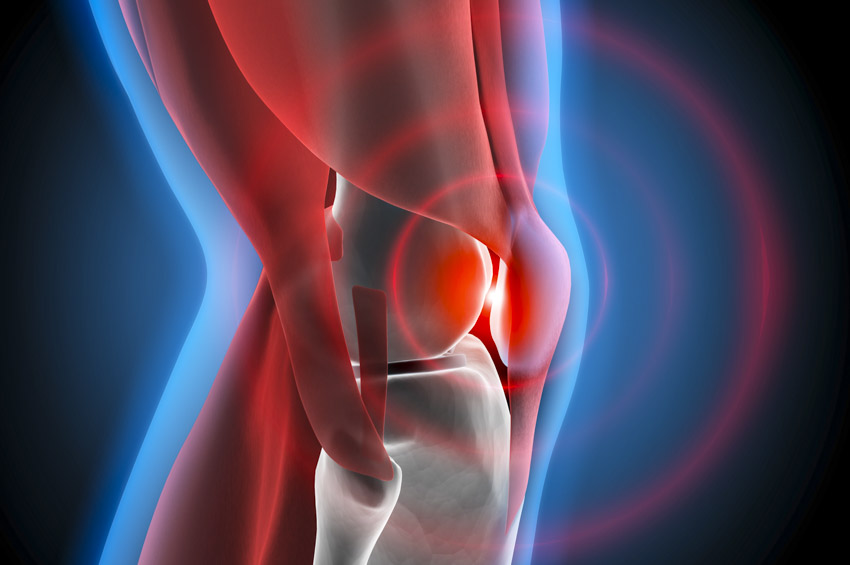
Knee osteoarthritis (KOA) often presents with pain, limited mobility, and a reduction in quality of life. Pathologically, it is characterized by changes in the articular cartilage, the subchondral bone, the intra-articular space, ligaments, and the articular capsule region. The researchers designed a laboratory experiment to determine the biochemical mechanisms by which acupuncture exerts its therapeutic actions. The randomized study of 30 experimental rabbits was divided into three arms: a control group (n=10), a knee osteoarthritis model group (n=10), and an acupuncture treatment group (n=10). For the KOA model group and treatment group, a KOA model was made by immobilization of the rabbit knee. Only the treatment group received warm needle acupuncture treatments. The control group did not receive any medical procedures or treatment for the duration of the study.
Results
The hospital researchers conclude, “In comparison with the control group, the expression levels of both TGF-beta1 [transforming growth factor beta 1] and IGF1 [insulin-like growth factor 1] were up-regulated in the KOA model group and then down-regulated after the warm-needling treatment.” They add that both microscopic and macroscopic improvements in the knee joint were observed. After the treatment, “the surface of knee cartilage became smoother.” Additionally, “less abnormal cell proliferations and clusters were found in the warm-needling group compared to those of the model group.” The research team determined that “acupuncture can inhibit the overexpression of TGF-beta1 and IGF-1 in the knee cartilage of rabbits with KOA, having a solid effect in improving pathological changes of knee cartilage.”
Acupuncture
Acupuncture treatment commenced one week after the KOA model was established. Acupuncture was applied once per day, for a total of four weeks. Needle retention time per acupuncture session was 20 minutes. Manual needle stimulation was applied to elicit a deqi response for each needle. Additionally, moxa cuttings were attached to each needle handle and ignited for 20 minutes to warm the needles. The acupoints used in the study were the following:
- EX-LE4 (Neixiyan, medial Xiyan)
- EX-LE5 (Waixiyan, lateral Xiyan)
- SP10 (Xuehai)
- GB34 (Yanglingquan)
The researchers note that Traditional Chinese Medicine (TCM) principles were used to make the acupoint selection for the study. This acupoint prescription is widely used on human patients. In Traditional Chinese Medicine, KOA is in the scope of Gu Bi (translated as bone impediment) and Xi Tong (translated as knee pain). The stagnation of external pernicious influences (e.g., wind, cold, damp, heat, phlegm) in the local meridians (acupuncture channels and collaterals) is a primary cause of KOA.
The TCM treatment principle is to invigorate local blood circulation and disperse stagnation. Neixiyan, Waixiyan, Xuehai, and Yanglingquan are located at the knee region. Needling these acupoints accelerates local blood circulation. According to the Huangdi Neijing (The Yellow Emperor’s Classic of Medicine), “the knee is the house of the tendons.” Yanglingquan (GB34) is the gathering point of the tendons. Needling this acupoint treats tendon issues, including those related to the knee joint. In this study, moxibustion was also applied as a part of warm needling therapy. Moxibustion warms the local area and supplements acupuncture to promote blood circulation and disperse stagnation.
Laboratory Detection
The researchers note, “KOA is caused by an imbalance between knee cartilage degradation and repair, the process of which is closely related to TGF-beta1 and IGF-1.” [1] This is supported by another research investigation, in which Stephen et al. concluded that “TGF-beta1 and IGF-1 plays an important role in cartilage homeostasis.” [2]
TGF-beta1 is a secreted protein involved in osteoblast formation and bone remodeling. There is a positive correlation between the concentration of TGF-beta1 and the degree of cartilage damage. Fahlgren et al. note that “high concentrations of TGF-beta1 in synovial lavage fluid seemed indicative for the later development of more severe OA changes in contrast to lower concentrations.” [3] Couchourel et al. document that “Elevated TGFβ1 levels in OA osteoblasts are responsible, in part, for the abnormal ratio of COL1A1 to COL1A2 and for the abnormal production of mature type I collagen. This abnormal COL1A1-to-COL1A2 ratio generates a matrix that blunts mineralization in OA osteoblasts.” [4] Homeostasis is the key. Tsai et al. find that “at proper levels, TGF-β1 could prevent OA from progression.” [5]
IGF-1 is a mitogen (substance that stimulates mitosis) that plays a role in regulating the proliferation, differentiation and apoptosis of chondrocytes. Lan et al. note that at the early stages of knee OA, normal increase in concentrations of IGF-1 activates bone growth and repair. [6] However, they also document that overexpression of IGF-1 leads to osteophyte formation and disease deterioration. In a related research, Wei et al. conclude, “TGF-1 regulates development and homeostasis of articular cartilage, and only TGF-1 at proper levels can accelerate cartilage repair and prevent OA’s progression.” [7]
Based on the findings, the Beijing University of Traditional Chinese Medicine research team concludes that “the relation between TGF-beta1 and IGF-1 levels and articular cartilage repair is a normal distribution.” They add, “the key of knee OA treatment is to reduce the overexpression of the two regulatory factors.” Their findings demonstrate that acupuncture stimulates a homeostatic response on expression of these factors.
Summary
Laboratory data indicates that acupuncture is effective for the treatment of KOA. According to the research covered in this article, common protocols involve the application of acupoints EX-LE4 (Neixiyan), EX-LE5 (Waixiyan), SP10 (Xuehai), and GB34 (Yanglingquan). Patients interested in learning more about treatment are recommended to contact local licensed acupuncturists.
Primary Research
[1] Gao L, Chen M, Yue P, Zhang R, Xin SC. Effect of Warm-needle Moxibustion on Expression of Transfer Growth Factor-beta1 and Insulin like Growth Factor 1 in Knee Cartilage of Rabbits with Knee Osteoarthritis [J]. Acupuncture Research, 2015,40(3):229-232.
Notes
[1] Gao L, Chen M, Yue P, Zhang R, Xin SC. Effect of Warm-needle Moxibustion on Expression of Transfer Growth Factor-beta1 and Insulin like Growth Factor 1 in Knee Cartilage of Rabbits with Knee Osteoarthritis [J]. Acupuncture Research, 2015,40(3):229-232.
[2] Trippel S B. Growth Factor Inhibition Potential Role in the Etiopathogenesis of Osteoarthritis [M]. Philadelphia: Lippincott Williams & Wilkins, 2004:47-52.
[3] Fahlgren A, Andersson B, Messner K. TGF-beta1 as a prognostic factor in the process of early osteoarthrosis in the rabbit knee. Osteoarthritis Cartilage. 2001; 9:195–202. doi: 10.1053/joca.2000.0376.
[4] Couchourel D, Aubry I, Delalandre A, et al. Altered Mineralization of Human Osteoarthritic Osteoblasts Is Attributable to Abnormal Type I Collagen Production. Arthritis and rheumatism. 2009;60(5):1438-1450. doi:10.1002/art.24489.
[5] Tsai S-H, Sheu M-T, Liang Y-C, Cheng H-T, Fang S-S, Chen C-H. TGF-β inhibits IL-1β-activated PAR-2 expression through multiple pathways in human primary synovial cells. Journal of Biomedical Science. 2009;16(1):97. doi:10.1186/1423-0127-16-97.
[6] Lan X, Liu XM, Ge BF et al. Observation of Serum IGF-1 Concentration of Primary Osteoarthritis in Guinea Pigs [J]. Orthopedic Journal of China, 2000, 7(7):667-668.
[7] Wei YX, Wei Z, Liu P et al. Experimental Study on the Repair of Articular Cartilage Defects and Prevention on Osteoarthritis by Intra-articular Injection of IGF-1, TGF-β1 [J]. Chinese Journal of Clinical Healthcare, 2010, 13(3):274-277.