Acupuncture regenerates neurons and promotes biological processes responsible for motor function recovery after spinal cord injuries. Often, motor function below spinal cord injuries deteriorates, which may lead to paralysis. Researchers discovered that acupuncture may prevent paralysis by stimulating repair. The researchers (Yang et al.) discovered that electroacupuncture produced important biological responses after a spinal cord injury necessary for the protection of motor neurons. Electroacupuncture increased the following:
- the total number of healthy neurons
- activity of acetylcholinesterase (AChE)
- the total number Nissl bodies
- glial cell line-derived neurotrophic factor (GDNF) in mRNA
The research demonstrates that electroacupuncture promotes repair of cells responsible for motor function, which may stimulate nerve repair sufficient for the prevention of paralysis after a spinal cord injury. One way to assess the degree of nerve injury is to assess the number of Nissl bodies or the number of neurons containing Nissl bodies, which are the sites of protein synthesis (Gulino and Gulisano). Another way to assess the degree of injury is through the activity of AChE, the enzyme necessary to regenerate neurons through the breakdown of the neurotransmitter acetylcholine (Nakamura).
Acupuncture has been proven to enhance AChE within the midbrain reticular formation (Ramey and Archer). This, coupled with additional research demonstrating that acupuncture is effective at reducing inflammation after an acute injury, indicates that acupuncture has a therapeutic role for spinal cord injury patients (Nayak et al.).
Plasticity is inherent in the central nervous system; therefore, regeneration is possible after a spinal cord injury (Bregman et al.). Treatment success is dependent upon the nervous system's ability to heal. Spinal motor neurons contain neurotrophic factors vital to the functioning of the neurons. Glial cell-derived neurotrophic factor (GDNF) is important for nerve function. GDNF improves the overall recovery of neurons and neuroglia by inhibiting cell death caused by injuries (Cheng et al., Bakshi et al., McCullough et al.).
In this controlled laboratory experiment, the thoracic segments of 60 healthy rats were compressed after humane anesthetization, resulting in moderate to severe spinal cord injuries. After compression, the lower extremities and torsos showed signs of paralysis, which is expected from a spinal cord injury. Next, the rats were divided into random control and electroacupuncture groups. The control group did not receive any treatment.
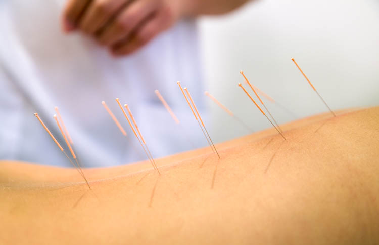
The electroacupuncture group began treatment 24 hours post-injury. Acupuncture points included Zusanli (ST36), Xuanzhong (GB39), Futu (ST32), and Sanyinjiao (SP6). The electroacupuncture device was an HB-EDT-II. These sessions were performed daily with a 30 minute needle retention time. Two groups of acupoints received electroacupuncture, alternately.
During weeks 2, 4, and 6, effects of electroacupuncture were assessed by removing four sections of spinal cord for examination under a microscope. Investigators looked for nerve swelling, capsular spacing, and vacuolar degeneration commonly seen in nerve damage, as well as Nissl bodies, GDNF, and AChE activity. In addition, the total number of motor neurons were compared between the control and electroacupuncture groups.
At 2 weeks, the control group demonstrated degeneration as would be expected following a spinal cord injury, including cell death and inflammation. However, in the electroacupuncture group, many neurons survived with only light swelling and necrosis. Moreover, there was a significant difference in the level of GDNF mRNA expression in the electroacupuncture group over the control. This demonstrates that electroacupuncture is effective at increasing GDNF expression in rats with spinal cord injuries.
At 4 weeks post-injury, rats in the electroacupuncture group showed large numbers of Nissl bodies, which stained larger and more darkly than the control group. As for the control group, the Nissl bodies were smaller in number and lightly stained. Eventually, in the control group, cellular edema and vacuolar degeneration increased and the Nissl bodies disappeared.
Remarkably, in the electroacupuncture group, Nissl body staining remained throughout 2, 4, and 6 week markers, and the number of motor neurons in the anterior horn of the spinal cord were significantly higher than in the control group. These findings suggest that electroacupuncture may promote the survival of motor neurons in the anterior horn of the spinal cord. AChE levels were also assessed throughout and it was observed that AChE activity in the motor neurons was lowest at 4 weeks after electroacupuncture and gradually increased toward the 6 week time period.
One surprising discovery was that electroacupuncture at Zusanli (ST 36) stimulated muscle contraction of the contralateral gastrocnemius, which suggests that electroacupuncture increases acetylcholine activity within the nerve itself through the stimulation of the acupoint. Yang et al. note that electroacupuncture generates sensory impulses in the deep tissues, which are transmitted afferently, activating neurons at the level of the spinal cord, resulting in muscle contraction.
Acupoints used in this study were correlated with spinal cord plasticity (Takeshige et al.). The current study shows the mechanisms of action and that electroacupuncture is specifically effective for the protection of neurons following a spinal cord injury. Compared with the control group that did not receive any treatment, the electroacupuncture group had an increase of AChE activity of the motor neurons in the anterior horn, which is vital for the successful treatment of the spinal cord after a spinal cord injury. Also, due to the greater number of Nissl bodies found in the electroacupuncture group, the study's findings suggest that electroacupuncture promotes the overall survival of motor neurons and enhances protein synthesis. This is the first major study to demonstrate the effect of acupuncture on GDNF mRNA expression in motor neurons of the anterior horn after a spinal cord injury. These findings and prior research demonstrate that electroacupuncture has a potentially important role for regaining motor function after a spinal cord injury.
References:
Yang J, Lv J, Wang H, Nie H (2015). Electroacupuncture promotes the recovery of motor neuron function in the anterior horn of the injured spinal cord. Neural Regeneration Research. (10)12:2033-2039.
Gulino R, Gulisano M (2013) Noggin and Sonic hedge hog are involved in compensatory changes within the motoneuron-depleted mouse spinal cord. J Neurospinal cord injury 332:102-109.
Nakamura M, Fujimura Y, Yato Y, Watanabe M, Yabe Y (1996) Changes in choline acetyltransferase activity and distribution following incomplete cervical spinal cord injury in the rat. J Neurospinal cord injuryence 75:481-494.
Nayak S, Matheis RJ, Agostinelli S, Shifleft SC (2001) The use of complementary and altemative therapies for chronic pain following spinal cord injury: a pilot survey. J Spinal Cord Med 24:54-62.
Bregman BS, McAtee M, Dai HN, kuhn PL (1997) Neurophic factors increase axonal growth after spinal coral injury and transplantation in the adult rat. Exp Neurol 148:475-494. Cheng H, Huang SS, Lin SM (2005)
Cheng H, Huang SS, Lin SM (2005) The neuroprotective effect of glial cell line-derived neurotrophic facror in fibrin in glue against chronic focal cerebral ischemia in conspinal cord injuryous rats. Brain Res 1033:28.
Bakshi A, Shimizu S, Keck CA, Cho S, Sean CB, David G, Morales D, Arenas E, Snyder EY, Watsn DT, Mclntosh TK (2006) Neural progenitor cells engineered to secrete GDNF show enhanced survival neuronal differentiation and improve cognitive function following traumatic brain injury. Eur J Neurospinal cord injury 23:2119.
McCullough MJ, Gyorkos AM, Spitsbergen TM (2013) Shopt-term exercise increase GDNF protein levels in the spinal cord of young and old rats. Neurospinal cord injuryence 240:258-268.
Takeshige C, Luo CP, Hishida F, Igarashi O (1990) Differentiation of acupuncture and nonacupuncture points by difference of associated opioids in the spinal cord in production of analysia by acupuncture and nonacupuncture point stimulation and relations between sodium and those opioids. Acupunct Electrother Res 15:193.
Ramer MS, Bisby MA (1998) Differences in sympathetic innervation of mouse DRG following proximal or distal nerve lesions. Exp Neurol 152:197-207.