CT scans reveal anatomical structures of acupuncture points.  A CT (computerized tomography) scan is a series of X-rays used to create cross-sectional images. In this study published in the Journal of Electron Spectroscopy and Related Phenomena, researchers used in-line phase contrast CT imaging with synchrotron radiation on both non-acupuncture points and acupuncture points. The CT scans revealed clear distinctions between the non-acupuncture point and acupuncture point anatomical structures.
Acupuncture points have a higher density of micro-vessels and contain a large amount of involuted microvascular structures. The non-acupuncture points did not exhibit these properties.
The researchers note that the state-of-the-art CT imaging techniques used in this study allow for improved three-dimensional (3D) imaging of a large field of view without artifacts. This greatly improves imaging of soft tissue and allowed the researchers to make this important discovery.
The acupuncture points ST36 (Zusanli) and ST37 (Shangjuxu) were shown to have very distinct structural differences than surrounding areas. At the acupuncture points, microvascular densities with bifurcations “can be clearly seen around thick blood vessels” but non-acupuncture point areas showed few thick blood vessels and none showed fine, high density structures. The acupuncture points contained fine structures with more large blood vessels that are several dozen micrometers in size plus beds of high density vascularization of vessels 15-50 micrometers in size. This structure was not found in non-acupuncture point areas.
The researchers note that the size of an acupuncture point “can be estimated by the diameter of microvascular aggregations….” They also commented that other research has found unique structures of acupuncture points and acupuncture meridians using MRI (magnetic resonance imaging), infrared imaging, LCD thermal photography, ultrasound and other CT imaging methods. The researchers commented that many studies using these technological approaches have already shown that acupuncture points exist. They note that “the high brightness, wide spectrum, high collimation, polarization and pulsed structure of synchrotron radiation” facilitated their discovery. They concluded, “Our results demonstrated again the existence of acupoints, and also show that the acupoints are special points in mammals.”
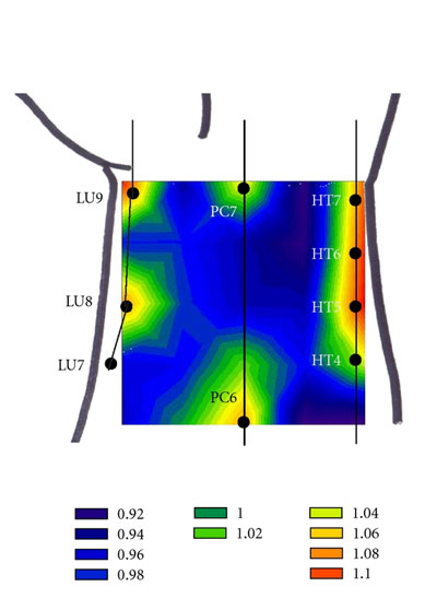
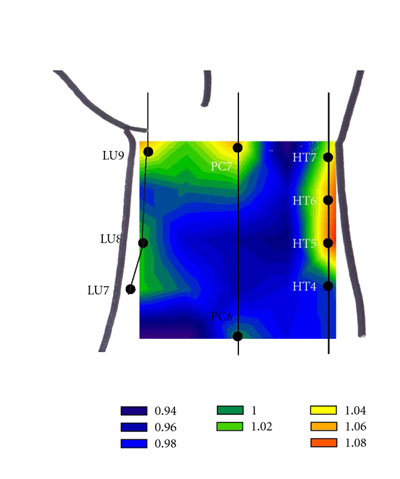
In another interesting study, researchers used an amperometric oxygen microsensor to detect partial oxygen pressure variations at different locations on the anterior aspect of the wrist. The researchers concluded that partial oxygen pressure is significantly higher at acupuncture points. Below are images from the study measuring the increase of partial oxygen pressure combined with an overlay of the local acupuncture point locations. The images map the Lung, Pericardium and Heart channels and their associated local points. Acupuncture points P7 and P6 clearly show high oxygen pressure levels as do the other acupuncture points in the region.
These measurements are not needled points but are natural resting states of acupuncture points absent stimulation. A truly unique finding, acupuncture points exhibit special oxygen characteristics. Acupuncture points and acupuncture channels are scientifically measurable phenomena in repeated experiments.
References:
Chenglin, Liu, Wang Xiaohu, Xu Hua, Liu Fang, Dang Ruishan, Zhang Dongming, Zhang Xinyi, Xie Honglan, and Xiao Tiqiao. "X-ray phase-contrast CT imaging of the acupoints based on synchrotron radiation." Journal of Electron Spectroscopy and Related Phenomena (2013).
Author Affiliations:
1. Liu Chenglin, Wang Xiaohua, Xu Hua; Physics Department of Yancheng Teachers’ College, Yancheng, China.
2. Liu Fang, Dang Ruishan; Anatomy Department of Second Military Medical University, Shanghai, China.
3. Zhang Dongming, Zhang Xinyi; Synchrotron Radiation Research Center of Fudan University, Shanghai, China.
4. Xie Honglan, Xiao Tiqiao; Shanghai Synchrotron Radiation Facility of Shanghai Institute of Applied Physics, Shanghai, China.
Minyoung Hong, Sarah S. Park, Yejin Ha, et al., “Heterogeneity of Skin Surface Oxygen Level of Wrist in Relation to Acupuncture Point,” Evidence-Based Complementary and Alternative Medicine, vol. 2012, Article ID 106762, 7 pages, 2012. doi:10.1155/2012/10a6762.
Yan X H, Zhang X Y, et al. Do acupuncture points exist? [J]. Physics in Medicine and Biology, 54 (2009):N143–N150.
Zhang Y, Yan X H, Liu C L, et al. Photoluminescence of acupuncture points “Waiqiu” in human superficial fascia [J]. J Lumin. 2006, 119-120:96-99.
Julia J. Tsuei, Scientific Evidence in Support of Acupuncture and Meridian Theory: I. Introduction. IEEE Engineering in Medicine and Biology Magazine. 1996, 15(3):58-63.
Li Lei, Yau To, Yau Chuen-heung. What Is the Origin of Acupoint. J. Acupunct. Tuina. Sci. 2012, 10 (2):125-127.
Song X J, Zhang D. Study on the manifestation of facial infrared thermography induced by acupuncturing Guangming (GB 37) and Hegu (LI 4) [J]. Chinese Acupuncture & Moxibustion. 2010, 30(1):51-54.
Liu P, Zhou G Y, Zhang Y, et al. The hybrid GLM‒ ICA investigation on the neural mechanism of acupoint ST36: An fMRI study [J]. Neuroscience Letters, 2010, 479: 267-271.
Fei L, Cheng H S, et al. The experimental exploration and the research prospects about the material basis and the functional characteristics of the meridian [J]. Chinese Science Bulletin, 1998, 439(6):658-672.