CT scan imaging reveals unique anatomical structures of acupuncture points. Published in Biomedical Engineering and Informatics, researchers used in-line phase contrast CT imaging with synchrotron radiation to capture images of acupuncture points.  They compared non-acupuncture point locations with real Traditional Chinese Medicine (TCM) acupuncture points. The CT scans reveal unique distinctions between the non-acupuncture points and acupuncture point structures.
State-of-the-art synchrotron radiation CT techniques allowed for improved soft tissue imaging and a larger field of view. The CT technological breakthrough led to the discovery of unique acupuncture point characteristics. A detailed look at acupuncture point ST37 (Shangjuxu) reveals a concentrated number of involuted microvascular structures. Non-acupuncture points lacked the dense concentration of microvascular vessels and involuted structures found in acupoints.
The researchers note that the in-line imaging techniques employed in the study improved the light path, enhanced the field of view and eliminated artifacts. The technological advancement allowed for a new look at the microstructures at ST37 and other points. The researchers note that acupuncture points have small microvascular structures and their bifurcations “can be clearly seen around the thick blood vessels.” In addition, acupuncture points demonstrate a dense number of fine microvascular structures. On the other hand, non-acupuncture points show thick blood vessels absent the unique, fine structures found in true acupuncture points. The researchers conclude, “Our results demonstrated again the existence of acupoints, and also show that the acupoints are special points in mammals.”
In a prior study published in the Journal of Electron Spectroscopy and Related Phenomena, the research team used in-line phase contrast CT imaging with synchrotron radiation on both non-acupuncture points and acupuncture points and made similar discoveries. The CT scans revealed clear distinctions between the non-acupuncture point and acupuncture point anatomical structures.
In an identical finding, acupuncture points had a higher density of micro-vessels and contained a large amount of involuted microvascular structures. The non-acupuncture points did not exhibit these properties. Similarly, the researchers noted that the advanced CT imaging techniques allowed for improved three-dimensional (3D) imaging of a large field of view without artifacts and enhanced soft tissue visualization.
Acupuncture points ST36 (Zusanli) and ST37 (Shangjuxu) demonstrated very distinct structural differences from surrounding areas. At the acupuncture points, microvascular densities with bifurcations “can be clearly seen around thick blood vessels” but non-acupuncture point areas showed few thick blood vessels and none showed fine, high density structures. The acupuncture points contained fine structures with more large blood vessels that are several dozen micrometers in size plus beds of high density vascularization of vessels 15 - 50 micrometers in size. This structure was not found in non-acupuncture point areas.
The researchers note that the size of an acupuncture point “can be estimated by the diameter of microvascular aggregations….” They also commented that other research has found unique structures of acupuncture points and acupuncture meridians using MRI (magnetic resonance imaging), infrared imaging, LCD thermal photography, ultrasound and other CT imaging methods. The researchers commented that many studies using these technological approaches have already shown that acupuncture points exist. They note that “the high brightness, wide spectrum, high collimation, polarization and pulsed structure of synchrotron radiation” facilitated their discovery.
In another interesting study, researchers used an amperometric oxygen microsensor to detect partial oxygen pressure variations at different locations on the anterior aspect of the wrist. The researchers concluded that partial oxygen pressure is significantly higher at acupuncture points. Below are images from the study measuring the increase of partial oxygen pressure combined with an overlay of the local acupuncture point locations. The images map the Lung, Pericardium and Heart channels and their associated local points. Acupuncture points P7 and P6 clearly show high oxygen pressure levels as do the other acupuncture points in the region.
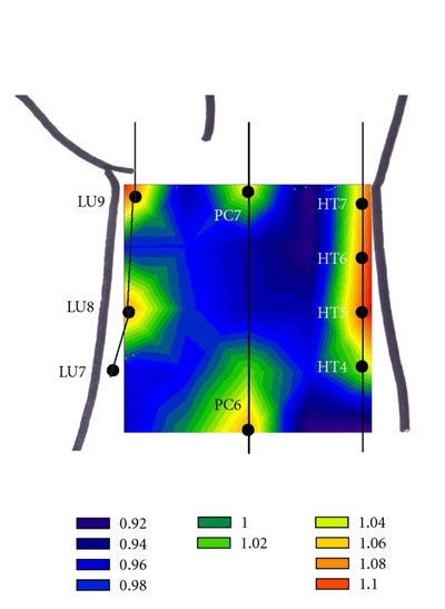
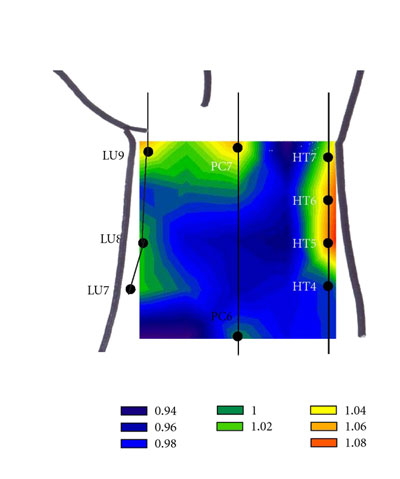
These measurements are not needled points but are natural resting states of acupuncture points absent stimulation. A truly unique finding, acupuncture points exhibit special oxygen characteristics. Acupuncture points and acupuncture channels are scientifically measurable phenomena in repeated experiments.
In related research, researchers have discovered an anatomical structure unique to acupuncture points. Wang, et. al., have identified a “vessel-like structure” made of “calcitonin gene related peptide (CGRP)-positive neurofibers in local tissues” at acupuncture points. The researchers discovered that “CGRP-positive nerve fibers were found to distribute in the dermis and subcutaneous layers of local tissues of acupoint ST 44, ST 36 and ST 32, mainly concentrating around the vessel-like structure.” They add, “CGRP-positive neurofibers are an important element in the local tissues of acupoint ST 44, ST 36 and ST 32 regions….” Miyauchi, et. al., note, “The calcitonin gene-related peptide (CGRP) plays important roles as a neurotransmitter/neuromodulator in the central nervous system, and as a potent vasodilator when secreted from peripheral, perivascular nerves through its specific receptors.” Wang, et. al,. used a laser confocal microscope to make the discovery of CGRP positive nerve fibers at acupuncture points.
CGRP is a type of neurotransmitter. Nerve fibers that are positive for the presence of CGRP play several roles in human physiology. For example, Hara-Irie, et. al., note that “CGRP-positive nerve fibers could be a crucial element in bone metabolism during bone growth and development.” Kunst, et. al., from the Yale School of Medicine (New Haven, Connecticut) note that CGRP is “a wake-promoting neuropeptide that regulates sleep maintenance at night.” Evans, et. al., from the University of Miami School of Medicine (Miami, Florida) note that CGRP is “a potent vasodilator neuropeptide.” The density of nerve fibers containing CGRP located at acupuncture points may correlate to the ability of acupuncture to stimulate signal conduction and induce health benefits.
Hongbao Ma of the Department of Medicine, Michigan State University (East Lansing) notes, “Calcitonin gene-related peptide (CGRP) is a 37 amino acid vasoactive neuropeptide that is widely distributed in central and peripheral nervous systems in mammals. CGRP was discovered in 1982 by molecular cloning of calcitonin (CT) gene.” Ma adds, “CGRP is secreted by primary afferents and causes primary hyperalgesia, and its expression increases in (the) dorsal horn under sensitization conditions. CGRP plays (an) important role in blood pressure system.” Given the discovery of CGRP in 1982, it is not unusual that the vessel-like physical structures of CGRP associated with acupuncture points have only recently been discovered.
Russell, et. al. note, “CGRP is a highly potent vasodilator and, partly as a consequence, possesses protective mechanisms that are important for physiological and pathological conditions involving the cardiovascular system and wound healing. CGRP is primarily released from sensory nerves and thus is implicated in pain pathways. The proven ability of CGRP antagonists to alleviate migraine has been of most interest in terms of drug development, and knowledge to date concerning this potential therapeutic area is discussed.”
Ling Zhao et. al., conclude that acupuncture is effective in the treatment of migraines and reduces pain intensity levels. Zhou et. al., find acupuncture effective in the prevention of migraines and links acupuncture’s therapeutic benefits to its ability to stimulate MLCK expression. The expression of myosin light-chain kinase (MLCK) is involved in the regulation of smooth muscle contraction. The researchers document a correlation between acute migraine attacks and decreases of MLCK via the CGRP signal system. The researchers discovered that applying acupuncture to acupoint GB20 (Fengchi) successfully upregulates MLCK expression and has “preventative and curative” effects for migraine patients.
In another investigation, Morry Silberstein, et. al., conclude that acupuncture points are related to both unmyelinated and myelinated afferent nerve fibers in a unique neuroanatomical structure not found in other areas of the body. The researchers used light microscopy on silver stained sections of acupuncture point P6 (silver stained human cadaver sample) and used confocal light microscopy on a live subject for acupuncture points GB20 and SP6. Control sites were compared with the acupuncture points.
At acupuncture points, it was discovered that a nerve bundle extended to the dermal-epidermal junctions. Each bundle branched into 2 sections perpendicular to each other. This anatomical phenomenon was not observed at the control sites. The researchers concluded that this acupoint neuroanatomical finding suggests that, “acupuncture may incise afferent unmyelinated axonal branch points, disrupting both neural transmission to the spinal cord and crosstalk along meridians, while simultaneously stimulating larger, myelinated afferents, thus explaining both the immediate and long-lasting effects of acupuncture.”
References:
Liu, Chenglin, Fei Wang, Xiaohua Wang, Fang Liu, Ruishan Dang, Dongming Zhang, Xinyi Zhang, Honglan Xie, and Tiqiao Xiao. "The micro-structure of Shangjuxu acupoint (ST37) by X-ray phase-contrast CT imaging based on synchrotron radiation." In Biomedical Engineering and Informatics (BMEI), 2014 7th International Conference on, pp. 85-89. IEEE, 2014. Author Affiliations:
1. Shanghai Synchrotron Radiation Facility, Shanghai Institute of Applied Physics.
2. Research Center of Synchrotron Radiation, Fudan University, Shanghai.
3. School of Physics and Electronics, Yancheng Teachers University.
4. Anatomy Department, Second Military Medical University, Shanghai.
Chenglin, Liu, Wang Xiaohu, Xu Hua, Liu Fang, Dang Ruishan, Zhang Dongming, Zhang Xinyi, Xie Honglan, and Xiao Tiqiao. "X-ray phase-contrast CT imaging of the acupoints based on synchrotron radiation." Journal of Electron Spectroscopy and Related Phenomena (2013).
Minyoung Hong, Sarah S. Park, Yejin Ha, et al., “Heterogeneity of Skin Surface Oxygen Level of Wrist in Relation to Acupuncture Point,” Evidence-Based Complementary and Alternative Medicine, vol. 2012, Article ID 106762, 7 pages, 2012. doi:10.1155/2012/10a6762.
Wang, C., W. J. Xie, M. Liu, J. Yan, J. L. Zhang, Z. Liu, and L. N. Guo. "[Distribution of calcitonin gene related peptide positive neurofibers in local skin tissues of" Neiting"(ST 44)," Zusanli"(ST 36) and" Futu"(ST 32) regions in the rat]." Zhen ci yan jiu= Acupuncture research/[Zhongguo yi xue ke xue yuan Yi xue qing bao yan jiu suo bian ji] 39, no. 5 (2014): 377-381.
Kunst, Michael, Michael E. Hughes, Davide Raccuglia, Mario Felix, Michael Li, Gregory Barnett, Janelle Duah, and Michael N. Nitabach. "Calcitonin Gene-Related Peptide Neurons Mediate Sleep-Specific Circadian Output in Drosophila." Current Biology (2014).
Ma, Hongbao. "Calcitonin gene-related peptide (CGRP)." Nat Sci 2 (2004): 41-47.
Russell, F. A., R. King, S-J. Smillie, X. Kodji, and S. D. Brain. "Calcitonin Gene-Related Peptide: Physiology and Pathophysiology." Physiological reviews 94, no. 4 (2014): 1099-1142.
Miyauchi, K., N. Tadotsu, T. Hayashi, Y. Ono, K. Tokoyoda, K. Tsujikawa, and H. Yamamoto. "Molecular cloning and characterization of mouse calcitonin gene-related peptide receptor." Neuropeptides 36, no. 1 (2002): 22-33.
Evans, Bornadata N., Mark I. Rosenblatt, Laila O. Mnayer, Kevin R. Oliver, and Ian M. Dickerson. "CGRP-RCP, a novel protein required for signal transduction at calcitonin gene-related peptide and adrenomedullin receptors." Journal of Biological Chemistry 275, no. 40 (2000): 31438-31443.
Hara-Irie, F., N. Amizuka, and H. Ozawa. "Immunohistochemical and ultrastructural localization of CGRP-positive nerve fibers at the epiphyseal trabecules facing the growth plate of rat femurs." Bone 18, no. 1 (1996): 29-39.
Zhao, Ling, Ji-xin Liu, Ying Li, Wei Qin, and Fan-rong Liang. "EFFECTS OF LONG-TERM ACUPUNCTURE TREATMENT ON RESTING-STATE BRAIN ACTIVITY IN MIGRAINE PATIENTS: A COMPARATIVE STUDY ON ACTIVE ACUPOINTS AND INACTIVE ACUPOINTS." Journal of Integrative Medicine 3 (2014): 234.
ZHOU Pei-juan, LI Bai, WANG Ai-cheng, LIU Chun-yan, WANG Yu, [Effect of Fengchi Point on the Expression of Myosin Light Chain Kinase on Middle Meningeal Artery of Migraine Model rats,] Acta Chinese Medicine and Pharmacology, 2014,(5), R285.5.
Morry Silberstein, Katharine Adcroft, Aston Wan, and Masimilliano Massi. Medical Acupuncture. Afferent Neural Branching at Human Acupuncture Points: Do Needles Stimulate or Inhibit?, doi:10.1089/acu.2011.0823. Department of Chemistry, Curtin University, Perth, Western Australia, Australia. Department of Pain Management, St. Vincent's Hospital Melbourne, Victoria, Fitzroy, Victoria, Australia.